 Search Articles

(CN) – It’s no sci-fi surgery robot, but brain cancer patients could soon find themselves in the operating room with an electronic doctor.
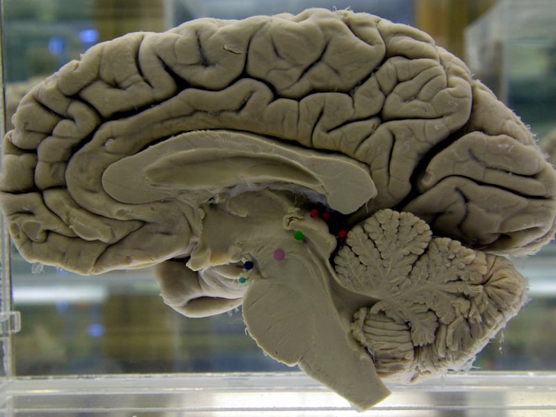
Physicians at the University of Michigan’s medical school are using artificial intelligence to diagnose brain tumors in real time.
Neurosurgeons Todd Hollon and Daniel Orringer described the breakthrough in a study published Monday in the journal Nature Medicine, with 35 colleagues as co-authors.
Hollon, the lead author, trained a convolutional neural network – a technology abbreviated as CNN that is better known for the warped and sometimes unsettling images created by Google’s Deep Dream image generator – on over 2.5 million images of patients’ brains.
The goal was to teach it to recognize the 10 most common types of brain cancer and to predict diagnoses with Michigan Medicine’s latest imaging tool – stimulated Raman histology, or SRH, a technology recently developed at the University of Michigan’s health system that rapidly produces images of tumor tissue.
“This is the first prospective trial evaluating the use of artificial intelligence in the operating room,” Hollon said in a statement issued by the university. “We have executed clinical translation of an AI-based workflow.”
After Hollon trained the network on SRH images from 415 different patients, he and his colleagues put it to work in the operating room. They tested the machine on 278 patients at three different institutions, comparing the CNN’s work to the more deliberately-paced work of a pathology lab, which requires a long wait for processing, staining and evaluating samples.
SRH cuts out the need for a pathology lab, according to the researchers, and with the help of the CNN pathologists can diagnose tumors within minutes at patients’ bedsides. That kind of efficiency means a lot in the operating room, where waiting on pathologist feedback can disrupt surgeons’ ability to make decisions on how to approach a case.
When that time is cut down to a few minutes, Hollon said, surgeons can get vital information sooner.
“It’s so quick that we can image many specimens from right by the patient’s bedside and better judge how successful we’ve been at removing the tumor,” he said.
The researchers describe the CNN as a “digital collaboration aid” to pathologists. With 94.6% accuracy compared to the 93.9% offered by conventional histology, the machine was able to correct pathologists’ mistakes and vice versa.
“It’s another way to help the pathologists and surgeons increase certainty while making important decisions in the operating room,” Hollon said.
The technology is some nine years in the making, according to Orringer, who began his work on SRH at the beginning of his tenure at Michigan Medicine as a neurosurgery resident and now also works with a manufacturer of the devices. His early work involved testing SRH technology on mice, and he was among those using the first SRH imager for neurosurgery, according to the university.
After all that work, widespread use of AI real-time diagnostics may be close at hand, Hollon said. The team at Michigan Medicine has already used the technology on over 500 patients, and Hollon predicts that it could be available for clinical use within three years.
“We’re transforming brain tumor diagnosis,” he said. “It’s a highly standardized tool that can deliver accurate diagnoses to a broad swath of brain tumor patients.”
Subscribe to our free newsletters
Our weekly newsletter Closing Arguments offers the latest about ongoing trials, major litigation and rulings in courthouses around the U.S. and the world, while the monthly Under the Lights dishes the legal dirt from Hollywood, sports, Big Tech and the arts.