 Search Articles

(CN) — In a major breakthrough for neuroscience over a decade in the making, an international team of researchers have completed a full brain map of an insect, the next step in what neuroscientists are hoping will become a better understanding of the mechanism of thought.
PublishedThursday in the journal Science , the brain map depicts the neural connections in the brain of a baby fruit fly, in higher definition than ever achieved before. The brain map, produced through a collaboration between the University of Cambridge and Johns Hopkins University, opens the door for a fuller understanding about how our brains are wired, and maybe even for the development of more sophisticated artificial intelligence systems.
“If we want to understand who we are and how we think, part of that is understanding the mechanism of thought,” said senior author Joshua Vogelstein, a Johns Hopkins biomedical engineer, in a statement. “And the key to that is knowing how neurons connect with each other.”
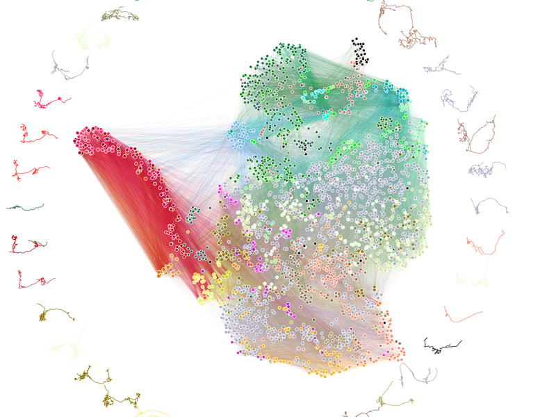
Neuroimaging in general has been used in medicine and brain research for many years, but the development of a highly detailed ‘connectome’ of an entire brain has required decades of work with complex technology.
The researchers’ connectome of a larva Drosophila melanogaster depicts 3,016 neurons and 548,000 connections between them. The team constructed the connectome by identifying the structure of the brain, locating individual neurons and tracing where they connect through synapses.
Although it seems unlikely, fruit fly brains are not dissimilar from human brains, especially in learning and decision making behaviors. In combination with its small size allowing for relatively quick imaging and mapping, the baby fruit fly became an ideal model organism for the project.
Even considering the fly’s relative small size, the project took 12 years to complete, as getting cellular-level imaging of the brain required slicing the brain into thousands of tissue samples, which then need to be imaged with electron microscopes, with the process taking as much as an entire day per neuron. From there, researchers needed to meticulously examine each neuron to reconstruct the pieces back into a full picture of the brain.
Cambridge took the helm in the electron microscope reconstruction, identifying each neuron and linking their synaptic connections.
Benjamin Pedigo, paper co-author and PhD candidate in Biomedical Engineering at Johns Hopkins said in an interview, “our co-authors were interested in finding meaningful groups of neurons with similar properties, which is often called cell typing in neuroscience. We used statistical models originally developed for networks — social networks, transportation networks, etc., — to place neurons into groups on the basis of their connectivity patterns.”
Researchers also found that the busiest circuits led to and away from the brain’s learning center.
Pedigo also said that the team noted that “a prevalence of ’non-cannonical’ connections, which don’t go from an axon to a dendrite (the textbook picture), but rather from an axon to an axon, dendrite to an axon, etc. These connections were known to exist but the prevalence of them in this brain was surprising. We also found that while a small number of neurons primarily receive inputs from one type of sensory information (e.g., visual, odor, etc), most of the brain has the potential to get input from a wide number of these signals.”
The code developed by Vogelstein and the John Hopkins Team is applicable to any other brain connection research, including adult fruit fly and mouse brain projects already in development. The team also noted that circuits they produced in their project had similarities to existing machine learning architectures.
This fruit fly connectome is the first complete map of an insect brain but partial and complete maps of other organisms have been explored since the 1970s. Until now, full connectomes have been produced only for species that have significantly fewer neurons in their bodies like the roundworm. Although partial connectomes of only tiny portions of the brain of more complex organisms, including humans, have also been generated, full connectomes are, according to researchers, unlikely to happen anytime soon.
“What we learned about code for fruit flies will have implications for the code for humans,” Vogelstein said. “That’s what we want to understand — how to write a program that leads to a human brain network.”
Subscribe to our free newsletters
Our weekly newsletter Closing Arguments offers the latest about ongoing trials, major litigation and rulings in courthouses around the U.S. and the world, while the monthly Under the Lights dishes the legal dirt from Hollywood, sports, Big Tech and the arts.