 Search Articles

(CN) — British scientists announced Monday that they have developed a machine-learning algorithm that can determine, with 98% accuracy, whether Alzheimer’s disease is present in a patient by looking at a single brain scan.
“Waiting for a diagnosis can be a horrible experience for patients and their families. If we could cut down the amount of time they have to wait, make diagnosis a simpler process, and reduce some of the uncertainty, that would help a great deal,” said Eric Aboagye, a professor of cancer pharmacology at Imperial College London and the study’s lead researcher, in a press release.
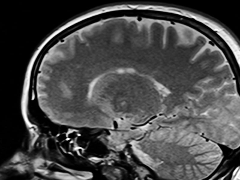
Many tests are used to diagnose Alzheimer’s, a kind of dementia that kills brain cells and deteriorates their connection, including brain scans made with a magnetic resonance imaging (MRI) machine.
“Although neuroradiologists already interpret MRI scans to help diagnose Alzheimer’s, there are likely to be features of the scans that aren’t visible, even to specialists,” said neurologist Paresh Mahotra, a researcher in ImperialCollege London’s department of brain sciences, in the release. “Using an algorithm able to select texture and subtle structural features in the brain that are affected by Alzheimer’s could really enhance the information we can gain from standard imaging techniques.”
The novelty of Aboagye’s team’s approach lies in adapting methods that were developed to classify cancer tumors to MRI scans of more than 400 patients with Alzheimer’s both early-onset and late-stage, healthy brains and also patients with Parkinson’s and other neurological conditions.
The data was provided by the Alzheimer’s Disease Neuroimaging Initiative, a cooperative, multisite study that aims to improve research on Alzheimer’s. The algorithm looked at each brain in terms of 115 regions, assessing each by such features as size, shape and texture.
The team “trained” the algorithm using these scans as inputs, teaching it the difference between regions and features that indicate Alzheimer’s disease and features that do not point to the presence of Alzheimer’s. By teaching it to tell signs from Alzheimer’s disease from red herrings, the algorithm became capable of making predictions when it was presented with new data.
Patients that were concurrently being tested for Alzheimer’s disease at Imperial College Healthcare NHS Trust in London, England, also offered their scans as data for the research. The scientists’ new approach was able to accurately predict whether a patient’s scan exhibited Alzheimer’s or not 98% of the time, and was even able to tell early from late-stage Alzheimer’s in 79% of patients.
The researchers tout their technique’s simplicity and claim it helps identify Alzheimer’s early in its development.
“Currently no other simple and widely available methods can predict Alzheimer’s disease with this level of accuracy, so our research is an important step forward,” Aboagye said. “Many patients who present with Alzheimer’s at memory clinics do also have other neurological conditions, but even within this group our system could pick out those patients who had Alzheimer’s from those who did not.”
Early diagnoses are difficult to ascertain with traditional methods, and while there exists no cure for the disease — though new treatments, even vaccines, may be reversing some of its consequences — patients with early notice are better able to receive support, prepare their loved ones and plan the rest of their lives with the condition in mind.
“Our new approach could also identify early-stage patients for clinical trials of new drug treatments or lifestyle changes, which is currently very hard to do,” Aboagye continued.
The team’s algorithm also made use of regions of the brain that were previously not examined to diagnose the disease. The cerebellum, which coordinates physical activity, and the ventral diencephalon, which figures in sight and hearing, are two such regions.
Aboagye’s research, published Monday in the peer-reviewed, open access journal Communications Medicine , was funded by a division of the United Kingdom’s National Institute for Health and Care Research Centre.
 Follow @cucumbermarg
Subscribe to our free newsletters
Our weekly newsletter Closing Arguments offers the latest about ongoing trials, major litigation and rulings in courthouses around the U.S. and the world, while the monthly Under the Lights dishes the legal dirt from Hollywood, sports, Big Tech and the arts.